Biology MECHANISM OF MUSCLE CONTRACTION

KEY TOPICS

`star` Effect of Action Potential
`star` Sliding Filament Theory
`star` Myoglobin in Muscles

EFFECT OF ACTION POTENTIAL

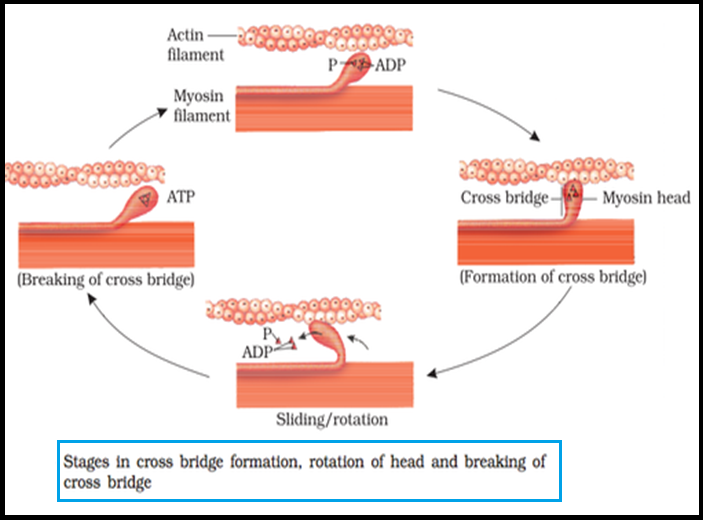
● `color{violet}("Mechanism of muscle")` contraction is best explained by the `color{brown}("sliding filament theory")` which states that contraction of a `color{violet}("muscle fibre")` takes place by the `color{violet}("sliding of the thin filaments")` over the thick filaments.

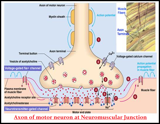
● `color{violet}("Muscle contraction")` is initiated by a signal sent by the central `color{violet}("nervous system (CNS)")` via a `color{brown}("motor neuron.")`

● A `color{violet}("motor neuron")` along with the `color{violet}("muscle fibres")` connected to it constitute a `color{brown}("motor unit.")`

● The junction between a `color{violet}("motor neuron")` and the `color{violet}("sarcolemma")` of the `color{violet}("muscle fibre")` is called the `color{brown}("neuromuscular junction")` or `color{brown}("motor-end plate.")`

● A `color{violet}("neural signal reaching")` this junction releases a `color{brown}("neurotransmitter (Acetyl choline)")` which generates an action potential in the `color{violet}("sarcolemma.")`

● This spreads through the `color{violet}("muscle fibre")` and causes the release of `color{brown}("calcium ions")` into the `color{violet}("sarcoplasm. ")`

SLIDING FILAMENT THEORY

● Increase in `color{violet}(Ca^(++))` level leads to the `color{violet}("binding of calcium")` with a subunit of troponin on actin filaments and thereby remove the `color{brown}("masking of active sites")` for `color{violet}("myosin.")`

● Utilising the energy from `color{violet}("ATP hydrolysis,")` the `color{violet}("myosin head ")` now binds to the exposed active sites on actin to form a `color{brown}("cross bridge.")`

● This pulls the attached `color{violet}("actin filaments")` towards the `color{brown}("centre of ‘A’ band.")`

● The`color{violet}(" ‘Z’ line")` attached to these actins are also `color{brown}("pulled inwards")` thereby causing a shortening of the sarcomere, `color{brown}("i.e., contraction.")`

● It is clear from the above steps, that during shortening of the `color{violet}("muscle")`, i.e., contraction, the `color{brown}("‘I’ bands get reduced,")` whereas the `color{brown}("‘A’ bands retain")` the length.

● The `color{violet}("myosin")`, releasing the `color{violet}("ADP and" P_1)` goes back to its relaxed state.

● A new `color{violet}("ATP binds")` and the `color{violet}("cross-bridge is broken.")`

● The ATP is again `color{violet}("hydrolysed")` by the `color{violet}("myosin head")` and the `color{violet}("cycle of cross bridge")` formation and breakage is repeated causing further sliding.

● The process continues till the `color{violet}(Ca^(++))` ions are `color{violet}("pumped back")` to the `color{brown}("sarcoplasmic cisternae")` resulting in the masking of `color{violet}("actin filaments.")`

● This causes the return of `color{violet}("‘Z’ lines")` back to their original position, i.e., `color{brown}("relaxation.")`

● The reaction time of the `color{violet}("fibres")` can vary in `color{violet}("different muscles.")`

● Repeated activation of the `color{violet}("muscles")` can lead to the `color{brown}("accumulation of lactic acid")` due to anaerobic `color{violet}("breakdown of glycogen")` in them, causing fatigue.

MYOGLOBIN IN MUSCLES

`star` `color{green}("Red Fibres:")`

● `color{violet}("Muscle")` contains a red coloured oxygen storing pigment called `color{brown}("myoglobin.")`

● `color{violet}("Myoglobin")` content is high in some of the `color{violet}("muscles")` which gives a reddish appearance.

● Such muscles are called the `color{brown}("Red fibres.")`

● These `color{violet}("muscles")` also contain `color{violet}("plenty of mitochondria")` which can utilise the large amount of oxygen stored in them for `color{violet}("ATP production.")`

● These muscles, therefore, can also be called `color{brown}("aerobic muscles.")`

`star` `color{green}("White Fibres:")`

● On the other hand, some of the `color{violet}("muscles possess")` very less quantity of `color{violet}("myoglobin")` and therefore, appear pale or `color{brown}("whitish.")`

● These are the `color{brown}("White fibres.")`

● Number of mitochondria are also few in them, but the amount of `color{violet}("sarcoplasmic reticulum ")`is high.

● They depend on `color{brown}("anaerobic process")` for `color{violet}("energy.")`


SiteLock